Vitreomacular Traction Syndrome
What is Vitreomacular Traction Syndrome?
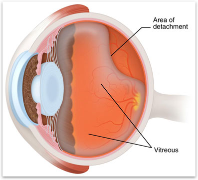
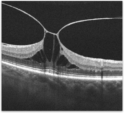
The macula is the special area at the center of the retina, which is responsible for clear, detailed vision. The macula normally lies flat against the back of the eye, like film lining the back of a camera. The vitreous is the clear gel-like substance that fills the interior of the eye (figure 1). As the vitreous naturally ages throughout life it shrinks and pulls away from the retina. In some people the vitreous can remain partially stuck to the macula. In these people the vitreous pulls on the surface of the macula and distorts the normal anatomy resulting in vitreomacular traction (figure 2).
What are the symptoms of Vitreomacular Traction Syndrome?
The condition is mild in most cases with very few symptoms. Since the macula is responsible for fine vision, blurriness is the most common symptom. As the macula is pulled on, it can begin to swell and in some cases a hole can develop in the macula. With more swelling, the central vision can become more blurry and distorted. If a hole develops, a small blind spot may develop in the center of the vision. In general, people do not go blind from vitreomacular traction. Even in the most severe cases, typically the peripheral or side vision is almost always maintained.
How is Vitreomacular Traction Syndrome diagnosed?
Your retina specialist can often detect vitreomacular traction syndrome by examining your retina using a dilated eye exam. A retinal scan called an Optical Coherence Tomography (OCT) is used to document the severity of the vitreomacular traction (figure 2). A photographic test called a fluorescein angiogram is useful in assessing if there is any damage to the surrounding blood vessels. For this test, fluorescein dye is injected into a vein. A series of photographs are taken of the retina as the dye passes through the back of the eye.
How is Vitreomacular Traction Syndrome treated?
Treatment is not necessary if symptoms are mild. When vitreomacular traction causes vision loss or the symptoms start to interfere with a persons normal daily activities, then surgery is recommended. In most cases, vitrectomy surgery is the most effective treatment to release the vitreomacular traction. During vitrectomy surgery, the vitreous is removed from the eye and the vitreous is separated from the back of the eye. If there is any scar tissue growing over the macula, micro-forceps can be used to gently remove the membrane. In very select patients, an injection of medication in the eye may cause the vitreomacular traction to release without the need for surgery.
What is the long-term impact of Vitreomacular Traction on my vision?
How much vision is restored after surgery for vitreomacular traction generally depends on its severity and how long it was present before surgery. Most patients note less distortion and are able to read two- to three more lines on the eye chart.

Figure 1. The macula is in the center of the retina. The vitreous gel fills up most of the inside of the eye.

Figure 2. OCT showing vitreomacular traction.

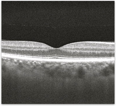
Figure 3. OCT showing the macula after release of the vitreomacular traction.

