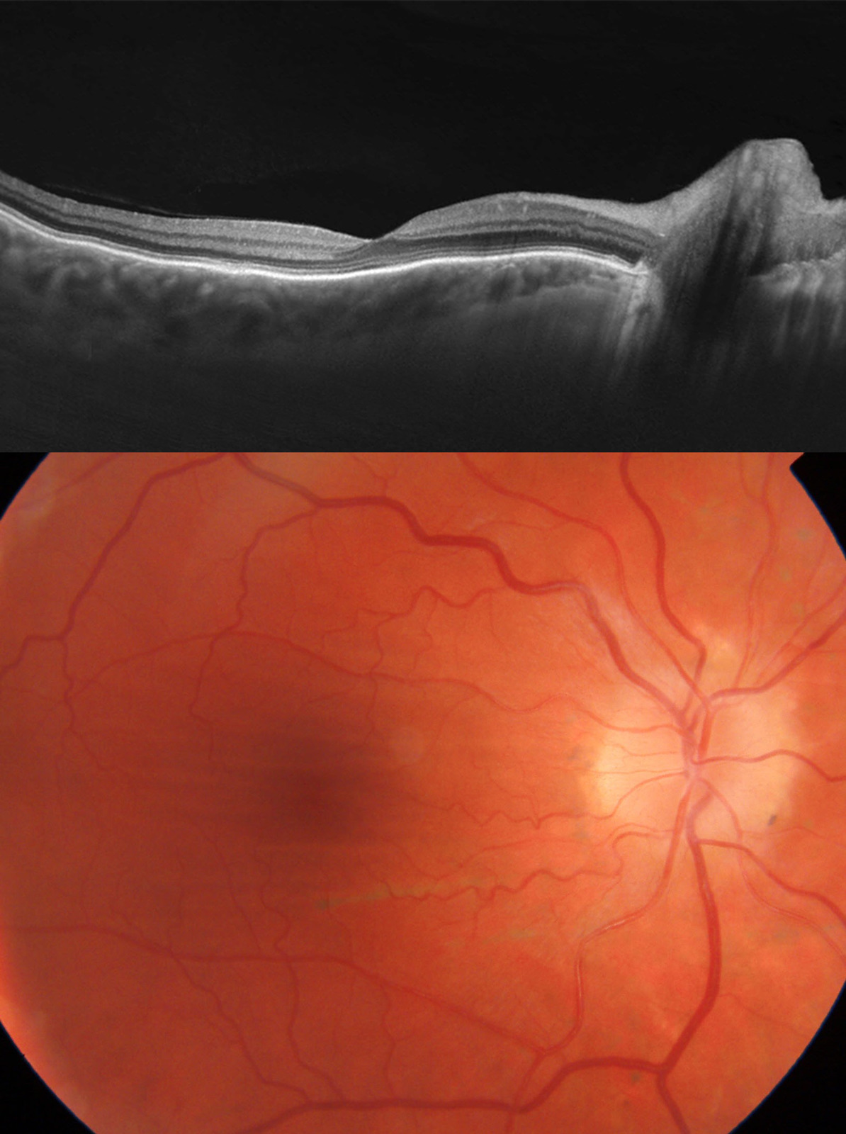
Image of the Month - June 2021

This image series demonstrates choroidal folds in a patient with optic nerve edema. Note the horizontal folds on the color image as well as RPE undulations seen on the OCT. The etiology for choroidal folds can be manifold including hyperopia, hypotony, scleritis, various posterior pushing mechanisms or oftentimes idiopathic. In conjunction with optic nerve edema, workup for any reasons that could contribute to posterior pressure on the globe including tumors or increased intracranial pressure is warranted.

