
Retinal Artery Occlusion
What is a Retinal Artery Occlusion?
The retina is a specialized layer of nerve tissue that coats the back of the eye and is responsible for helping you to see. A retinal artery occlusion occurs when blood flow in one of the arteries that feeds the retina is reduced or blocked. Because of the lack of oxygen in the retina, permanent severe vision loss can occur.
What causes a Retinal Artery Occlusion and who is at risk?
There are many different causes for a retinal artery occlusion. No single cause can explain the reason for every patient. The cause of a retinal artery occlusion may be discovered through a general medical evaluation. Sometimes, the source of a retinal artery occlusion cannot be identified, despite having a thorough examination and testing. Typically this condition occurs in individuals greater than 50 years old. The retinal artery is blocked most commonly because a small blood clot breaks off from around the heart or from a blood vessel in the neck and travels to the eye. The most common diseases that are associated with formation of a retinal artery occlusion include irregular heart beat (atrial fibrillation), narrowing of the arteries in the neck (carotid stenosis), heart disease, and blood clotting disorders.
Are there different types of Retinal Artery Occlusions?
There are two major categories of retinal artery occlusions, branch and central retinal artery occlusions. The arterial system supplying blood to the eye can be likened to a tree with one large trunk (central retinal artery) feeding smaller and smaller branches (branch retinal arteries). In a branch retinal artery occlusion (BRAO) a small branch of the retinal artery system becomes blocked (figure 1). In a central retinal artery occlusion (CRAO) the primary trunk of this is blocked (figure 2).
What are the symptoms of a Retinal Artery Occlusion?
Most patients experience a sudden painless loss of vision. In a CRAO the vision loss is usually very severe to the point where most people are not able to read any letters on the eye chart. The amount of vision loss in a BRAO depends on the size and location of the blockage. Typically only a part of the peripheral or central vision is affected in a BRAO.
How is a Retinal Artery Occlusion diagnosed?
Your retina specialist can diagnose an artery occlusion by examining your eye and using specialized testing to confirm the diagnosis and evaluate the extent of the blockage. A photographic dye test called a fluorescein angiogram can be used to identify the location of blockage and the extent of the loss of blood flow. Optical coherence tomography (OCT) is used to obtain a high-resolution anatomic scan of the retinal structure (figure 3). This technique is useful in documenting damage to the nerve tissue in the retina (figure 4).
What further testing needs to be done?
The most important aspect of your care is to try and identify the reason for the artery occlusion. It is possible that the underlying problem that caused the first artery occlusion could cause other artery occlusions in the other eye or elsewhere in the brain (a stroke). The actual tests that are ordered are different for every patient, however most patients need to have blood work, an echocardiogram (ultrasound of the heart) and a carotid Doppler (ultrasound of the vessels in the neck).
What are the complications of Retinal Artery Occlusions?
In severe cases of retinal artery occlusion where the retina does not get enough oxygen abnormal blood vessels can grow inside the eye (neovascularization). These new blood vessels are very problematic because they tend to bleed. In the worst cases bleeding can occur in the vitreous cavity or severe glaucoma can develop from the blood vessels growing near the front of the eye. Left untreated, these changes can result in irreversible blindness.
How is a Retinal Artery Occlusion treated?
The goal of treatment is to rapidly open the artery that has the blockage. Unfortunately the retina usually is irreversibly damaged when it does not get the oxygen and nutrition that it needs for just a short time. Many treatments have been described but there are really no good treatments for retinal artery occlusions. If the eye pressure is high, eye drops may be prescribed, since lowering the eye pressure sometimes helps relieve the arterial occlusion.
What is the prognosis?
Visual loss with CRAO is usually severe, with most people not able to read anything on the eye chart. In a small percentage of people, part of the central vision remains with good blood flow and a small central island of vision can remain. The visual field loss associated with a BRAO is usually permanent. If the part of the retina responsible for the central vision is unaffected the vision can remain good. In fact 80% of people with a BRAO recover vision good enough to drive!
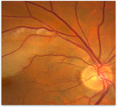
Figure 1. Branch Retinal Artery Occlusion (wedge shape of white retina). Note the cholesterol emboli in the artery at the tip of the Artery Occlusion.
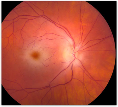
Figure 2. Central Retinal Artery Occlusion
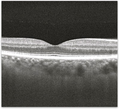
Figure 3. OCT of a normal macula
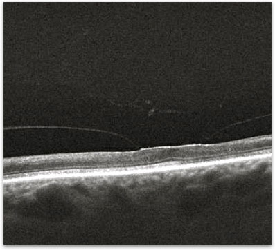
Figure 4. OCT showing the atrophy of the retina after having a Central Retinal Artery Occlusion