
Vitrectomy Surgery
What is a vitrectomy?
The retina is a specialized layer of nerve tissue that coats the back of the eye. A healthy retina is critical for normal vision. The vitreous is the clear gel-like substance that fills the interior of the eye. Vitrectomy is used to remove the vitreous gel from the eye, and is usually performed to allow treatment of disease of the retina and vitreous (figure 1). A Vitrectomy is always done by an ophthalmologist who has special training in treating diseases and conditions of the retina and vitreous.
How is a vitrectomy performed?
Vitrectomy is typically done in an operating room using local anesthesia. The surgery is performed using an operating microscope to allow a very detailed view of the retina. Tiny openings are created in the white part of the eye (sclera). Specialized micro-instruments are inserted into the vitreous gel through the openings in the sclera and the vitreous gel is cut out (figure 2). Other micro-instruments can be used to remove scar tissue and apply laser to the retina. Specialized saline, gas or silicone oil is injected into the eye to replace the vitreous gel at the end of the surgical procedure.
When is a vitrectomy recommended?
There are many different diseases that can be treated using vitrectomy. Some of the more common conditions that are treated include retinal detachments, diabetic retinopathy, macular hole, complications from cataract surgery and infections inside the eye.
How long will the vitrectomy take?
The length of the vitrectomy depends on the problem you have. Time for surgery can be from 30 minutes to over 3 hours. Your physician will discuss with you the approximate time he anticipates for your surgery.
What are some possible complications of vitrectomy surgery?
Most of the time people do well with vitrectomy surgery and there are no complications. However as with any surgery complications can occur. The most common include an increase in eye pressure, mild bleeding, drooping of the eyelid, dilated pupil, and double vision. A pre-existing cataract will progress more quickly. More serious but rare complications include retinal tears and detachments, infection and vision loss.
In Office Procedures
Intraocular InjectionLaser Surgery
Photodynamic Therapy
Pneumatic Retinopexy
Operating Room Procedures
Vitrectomy SurgeryScleral Buckle
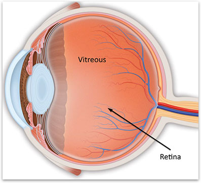
Figure 1. Anatomy of a normal eye showing the vitreous and the underlying retina at the back of the eye.
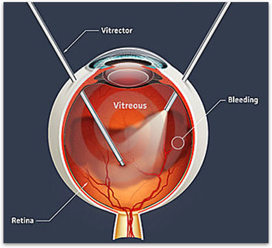
Figure 2. Typical set up for a vitrectomy involves the use of a light pipe and a suction cutter.
How much will my vision improve after surgery?
It is difficult to predict how much the vision will improve after surgery. Vitrectomy surgery is aimed at restoring the normal anatomy of the eye either by removing blood or debris from the eye, removing scar tissue from the eye or repairing a detached retina. The amount of vision improvement depends on how much damage the original disease has caused to the retina and how much your body is able to heal once the anatomy is repaired.